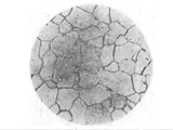
The microstructure of the Cr25Ni5Mo2N stainless steel is determined by examining the micrograph of a polished cross-section sampled after solution at 1030℃ followed by water quenching. It has been found that overall, the microstructure of the material is a bar-like along the long axis of the sample. The sample has been divided into four sections in order to analyze the microstructure.
The first section starts at a point directly adjacent to the edge of the sample and extends right up to the beginning of the second section. The different portions of the sample are identified by the numbers 1 through 4. It is clear that in the first section, there are some martensite laths. The martensite laths have an oval shape and stand out clearly against the background due to their perfectly defined boundaries. The laths are fine and their boundaries are well defined. The length of the laths is approximately 0.05mm.
In the second section, it is observed that there is a high degree of interlath disintegration. This is because the laths are still present, but are now beginning to form a network-like structure. The round laths remain clearly separated, with the large interlath gaps indicating the presence of many dislocations. The area fraction of the martensite is estimated to be around 10-15%.
The third section reveals a microstructure which is quite different from that of the previous sections. In this region of the microstructure, a clearly defined ferrite grain structure is observed. The size of the ferrite grain is relatively coarse and exhibits an equiaxed morphology. The area fraction of the ferrite is estimated to be around 40-45%.
The fourth section is different from the previous sections because it is composed of both an austenite and a ferrite microstructure. The austenite present in this section is coarse, and its area fraction is estimated to be about 15-20%. The ferrite occupies the remainder of the section, with an estimated area fraction of around 25-30%.
Overall, the microstructure of the Cr25Ni5Mo2N stainless steel sample is composed of both ferrite and martensite. The ferrite appears mainly as larger equiaxed grains, while the martensite appears mainly in the form of finely elongated laths. The area fraction of the ferrite and martensite is estimated to be about 40-45% and 10-15% respectively. The martensite laths are fine and have well-defined boundaries, while the ferrite grains are coarse and equiaxed. The austenite occupies a smaller area and its area fraction is estimated to be around 15-20%. The sample has been subject to solution at 1030℃ followed by water quenching, which has caused the formation of a network-like martensite structure.