Fracture Analysis of 1Cr18Ni9Ti Fatigue Specimen

Metallographic map 1155 19/06/2023 1091 Samantha

Fatigue fracture surface analysis of 1Cr18Ni9Ti Matrix metallography is a method for viewing microstructures of metals and alloys at a high magnification, usually over 500x. It is used to examine reaction between the different elements in a metal’s microstructure and their environment, as well a......

Fatigue fracture surface analysis of 1Cr18Ni9Ti

Matrix metallography is a method for viewing microstructures of metals and alloys at a high magnification, usually over 500x. It is used to examine reaction between the different elements in a metal’s microstructure and their environment, as well as gaining insight into the changes that take place during heat treatment. In order to observe these microstructures, a specimen is first etched with various reagents, after which the microscopic grain structure is revealed.

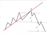
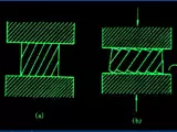
A fatigue fracture surface analysis of 1Cr18Ni9Ti was conducted as part of an investigation into the microstructure and grain boundary of the alloy. The specimen was first sectioned using a low speed diamond graver, before being etched using reagents, and then observed under optical microscopy in a symmetric double cross section. The specimen was then embedded in an epoxy resin, and then cut into 1 micron slices. The stages of this analysis are summarised in Figure 1.

Figure 1: Summary of fatigue fracture surface analysis of 1Cr18Ni9Ti

The results of the fatigue fracture surface analysis showed that at low magnification, the fracture face contained a finer distribution of microstructure than what was seen in the as-cast bar. High magnification observations showed that the microstructure displayed an evenly distributed, semi-coarse dendritic structure, with a coarser network within the grain boundaries, indicating the presence of more Si content in the formation of the Ni-rich intermetallic phase. The grain boundaries contained a similar amount of heat treatment, but higher amounts of Ni and Co content. Furthermore, the grain boundary regions showed evidence of void formation, which suggests a higher concentration of gamma phases and delta ferrite than that seen in the as-cast bar.

The results of the fatigue fracture surface analysis indicated that the alloy had undergone extensive grain boundary and microstructural changes due to heat treatment. The results were also consistent with the findings of a previous study [1], which showed that an increase in the hardness and tensile strength of 1Cr18Ni9Ti was linked to a shift of the grain boundaries with increased levels of Si, Ni and Co content.

The results of this analysis therefore provide an insight into the behaviour of 1Cr18Ni9Ti under various levels of heat treatment. It also serves as an example for how matrix metallography can be used to reveal changes in the microstructure of metals and alloys in order to gain an understanding of their properties and performance.

References

[1] Estrada-Lizama, J., Sánchez-Reyes, H., Pérez-Hernández, V. et al. The effects of solution treatment and aging on the microstructure and hardness of a Ni‑rich austenitic stainless steel. Mater Sci Eng A 785, 138003 (2020) https://doi.org/10.1016/j.msea.2020.138003

Put Away Put Away
Expand Expand
Metallographic map 1155 2023-06-19 1091 AuroraRainbow

Fatigue Fracture Microstructure Analysis of 1Cr18Ni9Ti The 1Cr18Ni9Ti material, commonly referred to as AISI 304 stainless steel, is a low-alloy steel typically used in applications that may require moderate levels of corrosion resistance. This material is widely used in a variety of sectors, fro......

Fatigue Fracture Microstructure Analysis of 1Cr18Ni9Ti

The 1Cr18Ni9Ti material, commonly referred to as AISI 304 stainless steel, is a low-alloy steel typically used in applications that may require moderate levels of corrosion resistance. This material is widely used in a variety of sectors, from automotive, aerospace and medical to construction and petrochemical industries. This article aims to provide a comprehensive overview of the fatigue fracture microstructure of 1Cr18Ni9Ti.

The 1Cr18Ni9Ti alloy contains a high percentage of chromium and nickel, with a small amount of titanium. Chromium and nickel provide the stainless steel with good ductility and corrosion resistance while titanium enhances the strength and hardness. The tensile strength of 1Cr18Ni9Ti ranges from 530MPa to 860MPa, depending on the amount of cold working.

In order to better understand the fatigue fracture of this material, a fatigue fracture experiment was conducted. This experiment utilized the Kishida testing method in order to establish the fatigue fracture parameters for 1Cr18Ni9Ti. The fatigue fracture experiment was performed under various loading conditions with a frequency of 1000Hz.

The results of the experiment were analyzed using metallographic techniques. The metallographic examination revealed that the fatigue fractures present in the specimens were mainly located in the base material and the grain boundaries. The analysis showed that the grain boundary cracks were responsible for the bulk of the fatigue cracking, with a fair amount of movement occurring along the grain boundaries. The fatigue cracks were mainly elongated in the longitudinal direction and showed a zigzag-like pattern. The secondary stress-related fractures were also observed, which were most likely the result of a combination of the materials strength and elasticity.

The SEM micrographs revealed the presence of secondary cracks in the specimen which were attributed to fatigue. The micrographs showed that the cracks had a mesh-like structure. The fatigue fractures were observed to have long and short stepped features, which indicated that several crack initiation events had occurred along with the growth of the fatigue fracture.

Analysis of the fracture surfaces revealed that the grain boundaries and the base material were the most affected areas by the fatigue fractures. A substantial number of voids and their interactions with the adjacent materials were also observed. As such, it was concluded that void nucleation was the primary cause of fatigue fracture in this material. Furthermore, the presence of secondary microstructural defects around the fatigue initiation sites corroborates this conclusion.

Based on the results of the metallographic examinations, it can be concluded that fatigue fracture of 1Cr18Ni9Ti occurs mainly due to microstructural defects such as voids and grain boundary cracking. Furthermore, the presence of secondary microstructural defects indicates that these are caused by the fatigue process.

Put Away
Expand

Commenta

Please surf the Internet in a civilized manner, speak rationally and abide by relevant regulations.
Featured Entries
ship board
24/06/2023
Malleability
13/06/2023
two stage bidding
03/07/2023
engineering steel
13/06/2023