Metallographic examination of a sample of heat treatment steel at 1100 °C x 20 min + 400 °C x 20s water cooling
Metallograph examination is an important part of testing to assess the microstructural changes in a material from a specific heat treatment. This paper explores the metallographic examinations of a sample of heat treatment steel at 1100 °C x 20 min followed by 400 °C x 20s water cooling to evaluate the presence of distinct microstructural features.
A sample of the heat-treated steel was obtained from a metallurgical lab in order to carry out the metallographic examination. The sample was sectioned using a reciprocating saw and roughly grounded using a dry grind wheel to bring out the microstructural features. The surface of the sample was then polished and lapped with various grinding and polishing solutions to generate a smooth and flat surface. On gaining a flat surface, the sample was mounted to a microscope and in order to investigate the microstructural features, optical and scanning electron microscopy was used.
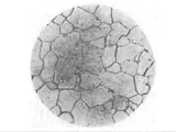
Optical microscopy revealed a very homogenous and uniform microstructure that displayed a fine and equiaxed grain structure. This suggests that the steel sample was heat treated at a temperature lower than the domain of full recrystallization. Additionally, adiabatic shear banding, associated with short‐time rapid cooling, was visible microscopically in the form of distinct fringes and lines superposed on the microstructure. Furthermore, no visible evidence of any carbide precipitation could be observed at the microstructural level.
Scanning electron microscopy (SEM) was then utilized to investigate the microstructure in more detail and it was observed that the microstructure was more complex at certain areas. These areas displayed the presence of both ferrite and austenite grains. The grain boundaries were mostly of the low-angle variant which is indicative of a small grain size. The rimming of ferrite with austenite was also present in small amounts. Additionally, the presence of shattered arches and dimples in the austenite ahead of ferrite could be observed which indirectly suggested that the observed microstructure was due to mechanical strain rather than a thermal process.
Finally, from the metallographic examination of the sample, it could be concluded that the heat treatment process of holding the sample at 1100 °C x 20 min followed by 400 °C x 20s water cooling resulted in the formation of a fine-grain structure in the steel. The presence of fractured arches and dimples in the austenite ahead of the ferrite grains is indicative of a mechanical strain as the cause of the observed microstructure. These findings are in line with that of the optical microscopy examination.