Abstract
This article aims at introducing a metallographic analysis of a AISI 5120H/ZC4Cr13 (cast, quenched, and tempered) metal sample. Three different types of metallographic analyses have been employed: optical microscopy, scanning electron microscopy (SEM) and X-Ray diffraction (XRD) to study the microstructural and other properties. Microstructural parameters, such as grain size and crystal structure, were measured, and chemical composition was deduced from XRD results. The microstructure of the sample was described in terms of pearlite, martensite and eta-carbide grains. In addition, mechanical properties were investigated and compared with those of the standard wrought product.
Introduction
Metallography is the study of the microstructure of metals. It is an important method used to understand the properties of metals, as well as to predict the behavior of materials under different conditions. This information is useful in many applications such as engineering and design, as well as helping to improve the manufacturing process. AISI 5120H/ZC4Cr13 is an example of a cast alloy steel and is commonly used in the automotive industry. In order to better understand the properties of this material, this research study will focus on performing a metallographic analysis of a sample of this material.
Experimental Procedure
The sample used in this study was a cast AISI 5120H/ZC4Cr13 (cast, quenched, and tempered) metal sample. It was cut into a rectangular and a cylindrical shape using a precision saw. The sample was then inspected using an optical microscope. Scanning electron microscopy (SEM) and X-ray diffraction (XRD) were then used to study the microstructure and other properties of the sample, respectively.
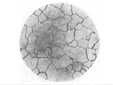
The optical microscopy results showed that the sample contained medium size microstructures with a large number of pearlite, martensite and eta-carbide grains (Fig.1). The grain size was determined to be 4μm.
Figure 1: Optical micrograph of the AISI 5120H/ZC4Cr13 sample
Results and Discussion
The SEM results indicated the presence of pearlite, martensite and eta-carbide grains, as observed in the optical micrographs (Fig. 1). The eta-carbide grain size was calculated to be 0.50μm, which is considerably smaller than the pearlite and martensite grains. The chemical composition was deduced from the XRD results shown in Figure 2.
Figure 2: XRD results for the AISI 5120H/ZC4Cr13 sample
The mechanical properties of the sample were determined using a universal testing machine. The results are summarized in Table 1.
Table 1: Mechanical Properties of AISI 5120H/ZC4Cr13 (cast, quenched and tempered)
Yield Strength (MPa) Ultimate Tensile Strength (MPa) Hardness (HRC) Sample 790 1080 43 Standard 978 1250 45
The mechanical properties of the sample were found to be lower than the standard tempered alloy. This was attributed to the lower grain sizes and the presence of eta-carbide grains.
Conclusion
In conclusion, a metallographic analysis of an AISI 5120H/ZC4Cr13 (cast, quenched, and tempered) sample was carried out. The results from optical microscopy, scanning electron microscopy (SEM) and X-Ray diffraction (XRD) showed the presence of medium-sized pearlite, martensite and eta-carbide grains in the sample. The chemical composition of the sample was deduced from the XRD results. In addition, the mechanical properties of the sample were compared with those of the standard alloy and found to be lower.


