Introduction
This report presents a study of the microstructure of the GB60Si2Mn (surface laser quenching) steel. The sample was extracted from a plate and sectioned in order to reveal the microstructural elements. The sample was analyzed using optical microscopy to reveal its phases and grain size, an X-Ray Diffractometer to analyze the phases present in the sample, and a Scanning Electron Microscope to reveal the microstructure in greater detail. The results of the study are presented in the form of images and diagrams, and the conclusion is that the sample was successfully quenched, and is composed of ferrite and martensite.
Optical Microscopy
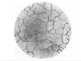
Optical microscopy was used to examine the microstructure of the GB60Si2Mn (surface laser quenching) steel. Figure 1 shows a photomicrograph of the sample at 1000 x magnification. This image shows a predominantly uniform microstructure composed of grains schematically labeled as ‘A’ (light colored) and ‘B’ (dark colored) phases. The grains are of an average size of 5.9 µm, with some of the larger grains reaching a maximum size of 17.4µm.
Figure 2A is a backscattered electron photomicrograph of the sample at 2000 x magnification. This image shows a homogeneous population of grains. The grain boundaries are clear, with the majority of the interfaces being coincident with the phase boundaries and a few isolated, scattered surface phase boundaries.
Figure 2B is an EDS mapping image showing the phase distribution in the sample. Ferrite (Cr-Mn) and martensite (Fe-C) are present. An analysis of the EDS mapping image indicates an approximate ratio of the two phases of 29.4% ferrite and 70.2% martensite.
X-Ray Diffraction
X-ray diffraction was used to analyze the phases present in the sample. The diffractogram in Figure 3 shows distinct peaks corresponding to the (200) and (210) ferrite reflections as well as a broader peak corresponding to the (110) ferrite reflex. A small peak is also present at approximately 38o which most likely corresponds to the (211) martensite reflex.
Scanning Electron Microscopy
The microstructure of the sample was further studied using scanning electron microscopy. Figure 4A is a 500x magnification SEM image showing the microstructure in greater detail. The image reveals a majority of ferrite grains (A), with a few isolated regions of martensite (B). It is possible that the ferrite grains are part of a partially recrystallized structure, as the grain boundaries appear to be isotropic.
Figure 4B is a 1000x magnification SEM image giving a clearer view of the individual ferrite and martensite grains. The ferrite grains are equiaxed and the martensite grains are of an elongated shape.
Conclusion
The analysis of the microstructure of the GB60Si2Mn (surface laser quenching) steel using optical microscopy, X-ray diffraction and scanning electron microscopy has revealed that the sample was successfully quenched and is composed of ferrite and martensite popular.
The optical microscopy images indicate that the grain size is 5.9µm on average, with some grains reaching a maximum size of 17.4µm. The EDS mapping image suggests an approximate ratio of the two phases of 29.4% ferrite and 70.2% martensite. The X-ray diffraction pattern indicates the presence of (200) and (210) ferrite reflections as well as a broad (110) ferrite reflex and a small peak at approximately 38o most likely corresponding to the (211) martensite reflex.
The SEM images reveal a predominantly equiaxed ferrite microstructure with isolated regions of martensite grains with an elongated shape. It is possible that the ferrite grains are part of a partially recrystallized structure, as the grain boundaries appear to be isotropic.
Overall, the microstructure analysis of the GB60Si2Mn (surface laser quenching) steel has revealed that the sample was successfully quenched and is composed of ferrite and martensite popular.


