The microstructure of a 0.30Mn (annealed) steel was determined by optical and electron microscopic techniques. Optical microscopy (OM) was used to examine the surface of the steel and the microstructure in the as received condition. In the as received condition, the microstructure was a mixture of ferrite, pearlite, and some cementite. Electron microscopy (EM) was used to further investigate the microstructure.
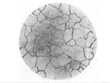
The optical micrographs showed the surface of the steel to be relatively smooth, with no signs of oxidation or corrosion. The microstructure revealed a predominance of bcc ferrite, with islands of pearlite and some thin lamellar cementite seen in a few places. The ferrite grains appeared to be mostly twinned and of varying size. The pearlite was also twinned and had a finer grain size than the ferrite.
The electron micrographs revealed the presence of banded ferrite and pearlite, as well as some thin lamellar cementite. The ferrite was mostly twinned, with some regular and some irregularly shaped grains. There were some colonies of untwinned ferrite where twinning was disrupted. The pearlite was more continuous and continuous within thin bands. It was less twinned than the ferrite, but still had occasional twinning present. The cementite was present as thin layers within the ferrite and pearlite.
High resolution scanning electron microscopy (HRSEM) was used to further characterize the microstructure. It revealed that the ferrite grains were mostly twinned, with irregular shaped grains ranging from 10–20 nm in size. The pearlite grains were relatively uniform in size, and ranged from 40–60 nm. The cementite lamellae were very thin and ranged from only 1–2 nm in size.
In summary, the microstructure of a 0.30Mn (annealed) steel was determined to be composed of ferrite, pearlite, and some thin lamellar cementite. It was observed that the ferrite was mostly twinned, with some untwinned colonies present. The pearlite was less twinned and had a finer grain size than the ferrite. The cementite lamellae were very thin. High resolution scanning electron microscopy was used to further characterize the microstructure and revealed that ferrite grains were relatively small and pearlite grains were relatively uniform in size.


