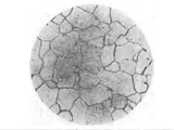
Optical microscopy is an important tool for observing and studying the microstructure of substances. It provides magnified images from microscopic sizes to macroscopic sizes that can be manipulated in various ways. Optical microscopes are divided into two categories: light microscopy and electron microscopy.
Light microscopy:
Light microscopy is the most widely used type of optical microscopy, providing images of good optical quality, greater depth of field, and relatively high speed imaging. Light microscopes work by using visible light, which can be generated by various sources, and then focusing it through a series of lenses to create an enlarged image. Light microscopes can be used to observe objects as small as 0.2 micrometers and as large as several centimeters.
The most common type of light microscopy is brightfield microscopy, which simply uses bright light as the source and is ideal for examining transparent or colorless specimens. Brightfield microscopy is easy to set up and uses a wide range of illumination sources, from tungsten, halogen, and quartz-halogen lamps to LED and fluorescent light sources.
Other types of light microscopy include phase contrast microscopy (which is used to observe transparent specimens), darkfield microscopy (which is used to observe very small or transparent objects), and polarization microscopy (which is used to study optical properties of a specimen). In addition, there are also special types of light microscopy, such as fluorescence and confocal microscopy, which are used to study samples labeled with fluorescent markers.
Electron microscopy:
In electron microscopy, an electron beam is used as the source of illumination instead of visible light, providing images of much higher resolution. Electron microscopes are divided into two categories: scanning electron microscopes (SEMs) and transmission electron microscopes (TEMs).
In a scanning electron microscope (SEM), an electron beam scans a specimen and collects secondary electrons that are emitted by the specimen. These secondary electrons are then used to produce a topographical image. This type of microscope is used to observe three-dimensional features of specimens, such as the electrically conducting surfaces of metal films or semiconductor devices.
In a transmission electron microscope (TEM), a thin sample is illuminated with a finely focused beam of electrons, which penetrate the sample and then produce a magnified image. This type of microscope produces very high-resolution images and can be used to observe objects as small as 0.1 nanometers.
Conclusion:
Optical microscopy is a powerful tool for observing and studying the microstructure of substances. It is divided into two categories: light microscopy and electron microscopy. Light microscopy is used for observing transparent or colorless specimens, while electron microscopy is used for producing very high-resolution images. Both types of microscopes can be used to observe objects as small as 0.2 micrometers, and each has its advantages and disadvantages.


